IMU Arrays: The Biomechanics of Baseball Pitching
Eric Berkson MD, Ryan Aylward MS, James Zachazewski DPT, ATC, Joseph Paradiso PhD, Thomas J. Gill MD
Massachusetts General Hospital
Abstract
Previous biomechanical studies have attempted to quantify
the mechanics of throwing and to measure the forces
sustained in the upper extremity during high-velocity pitching.
Biomechanical testing of pitchers in its current state, however,
is subject to inaccuracy and cumbersome to perform. Testing
requires controlled laboratory conditions where “high-speed”
cameras are set in fixed positions around the subject, and the
motion of the arm is tracked with navigational markers affixed
to the pitcher. Variables such as acceleration and velocity are
derived from a series of calculations based on positional data.
We hypothesized that direct measurements of acceleration and
velocity could improve kinematic analysis of baseball pitching.
To assess this hypothesis, a controlled validation study of a
novel Inertial Measurement Unit (IMU) array was performed.
Each IMU consists of three-dimensional accelerometers and
gyroscopes, permitting direct measurements of acceleration and
angular velocity. Simultaneous testing of a single professional
baseball pitcher was undertaken utilizing both “high-speed”
camera-based motion tracking and our newly developed IMU
array. Results indicated an acceleration phase during the pitching
cycle lasting 0.022 seconds. During this phase, traditional
motion tracking cameras recorded four data points. Thirty data
points were recorded by each IMU. The IMUs recorded 60.4g’s
of acceleration at the shoulder and 83.0g’s of acceleration at the
wrist. Acceleration over 100g’s was documented at the hand.
While no statistical comparison between systems was possible
in this early proof-of-concept study, the IMU array successfully
recorded appropriate rises in acceleration and velocity when
compared to the camera-based motion-analysis system and
offers the first direct measurement of acceleration in a professional
pitcher. As such, the IMU array promises to provide more
accurate kinematic measurements than alternative methods.
The technique also allows measurements outside of controlled
laboratory conditions and therefore could provide positive
practical and clinical applications ranging from improved player
training to injury prevention and rehabilitation.
Introduction
Pitching injuries have increased markedly over the past
few decades. In 1973, Tullos noted that 50% of professional
baseball pitchers at some point in their career experience elbow
or shoulder pain sufficient enough to keep them from throwing.
1 Between 1989 and 1999, the number of pitchers who
became disabled increased 54% and the number of days missed
by pitchers increased by 58%.2 This prevalence of injuries is
not limited to professional athletes. Andrews examined 5-year
periods within his practice and, comparing the two most recent
five-year periods, noted twice as many elbow surgeries for professional
pitchers, four times as many for collegiate pitchers,
and six times as many for high school pitchers.3
Most pitching injuries are a result of repetitive microtrauma
where repeated submaximal loads result in cumulative
microtrauma to the soft tissues over time.1, 4-7 These overuse
injuries have been attributed to many factors including pitch
counts, pitch types, pitch mechanics, physical conditioning,
periodization, nutrition, and supplements. Changes due to
improper mechanics, poor dynamic stability or muscle fatigue
negatively influence performance and may increase vulnerability
to injury.8, 9 An understanding of the biomechanics of
baseball pitching can assist in minimizing potential for injury,
preventing overuse injuries, and evaluating rehabilitation protocols.
Previous biomechanical studies have attempted to characterize
and quantify the mechanics of pitching and to
understand the relationship between factors leading to injuries.
Initial studies in the 60’s and 70’s were performed with
visual examinations of stroboscopic images.1 As technology has
advanced, improved mechanisms of analysis have permitted
more quantitative analysis. Today, computerized motion-tracking
cameras are used to follow markers placed on the subject.
Three-dimensional positional data of each body part is derived
and velocity, acceleration, and forces calculated secondarily.
Werner, Hawkins, and Gill utilized three 120 hertz cameras and
video analysis to determine positional data.10, 11 Andrews and
the American Sports Medicine Institute (ASMI) utilized six 240
hertz motion tracking cameras to perform these same calculations.
3, 4, 8, 12-14
Computerized tracking of the upper extremity, however,
remains challenging. Numerous markers are affixed to the
body, while multiple cameras are used to ensure that each
marker is always in view. Ideal environments are required to
ensure proper visualization by the cameras. For this reason,
a specialized laboratory is usually required for accurate data
capture. Although real-size pitching mounds have been built
within these environments and playing conditions have been
simulated, the constraints around data capture prevent direct
application to live practice and game activities.
These analyses have also been challenging due to the
magnitude of the velocities and accelerations involved. The
acceleration phase of pitching lasts approximately 20-40 milliseconds.
During this time, elbow extension velocities have been
calculated between 2,500 and 4,500 deg/sec.10, 13, 15, 16 Internal
rotation of the shoulder has been approximated at 10,000
deg/sec.10, 14 Accelerations of the arm, derived from the second
derivative of the positional data, are estimated to be between
300,000 deg/sec2 and 500,000 deg/sec2.16 At these extreme
speeds and with analysis only at 240 hertz, only 4-6 points of
measurements of the arm are possible, and smoothing errors
are introduced with each level of analysis. With so few data
points, calculations of acceleration simply exceed the resolution
of these motion-tracking cameras.
The purpose of this proof-of-concept study was to develop
and validate a new method of motion analysis that would not
suffer from the same level of environmental and measurement
constraints. During this study, a portable and wearable set of
accelerometers and gyroscopes were used to directly measure
velocity and acceleration about the elbow and shoulder during
a baseball pitch. Over the past decade, accelerometers and
gyroscopes have increased in accuracy and decreased in size
and cost. These devices, which respectively measure acceleration
and angular velocity, have been made small enough to
become practical in real-world applications.17 Accelerometers
outside of medicine have been used to detect a falling laptop
(ThinkPad Technologies) or to sense if airbag deployment is
necessary. More recently, accelerometers have been used to
measure lower extremity joint angles over time.18-21 Wearable
devices, with units on the leg and thigh, have been used to
track activity levels of subjects wearing the apparatus.17, 22, 23 In
addition, wireless three-dimensional accelerometers combined
with gyroscopes (Inertial Measurement Units — IMUs) have
recently been utilized to measure inertia for gesture recognition
by the MIT Media Lab.24, 25
 For our study, we hypothesized that a portable IMU array
could provide a direct measurement of the kinematics of baseball
pitching at least as accurate as the current standard of
camera-based tracking motion analysis. To assess this hypothesis,
a controlled validation study evaluated the kinematics of
a professional baseball pitcher, comparing our newly developed
array of accelerometers and gyroscopes to the traditional “highspeed”
tracking cameras.
For our study, we hypothesized that a portable IMU array
could provide a direct measurement of the kinematics of baseball
pitching at least as accurate as the current standard of
camera-based tracking motion analysis. To assess this hypothesis,
a controlled validation study evaluated the kinematics of
a professional baseball pitcher, comparing our newly developed
array of accelerometers and gyroscopes to the traditional “highspeed”
tracking cameras.
METHODS
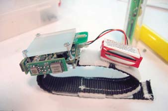
A pilot study of a prototype IMU array was completed
during spring training 2006. After IRB approval and informed
consent and under the direction of the subject’s coach, a single
professional baseball player underwent simultaneous biomechanical
testing utilizing both a camera-based motion tracking
system and our newly developed IMU array (Figure 1).
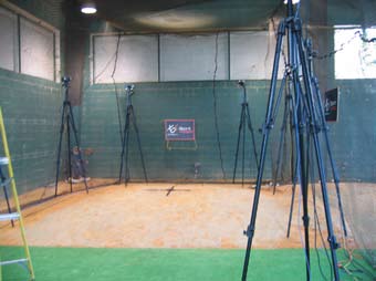
A camera-based motion analysis system (XOS
Technologies), employing “high-speed” cameras operating at
180 Hz, allowed positional tracking of each pitch. A series of
10 motion analysis cameras were set-up on a regulation-sized
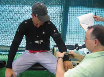
pitching mound (Figure 2). Subjects were fit with both passive
electrodes for the camera-based motion analysis and a 6
segment wireless IMU array (Figure 3). Inertial measurement
units were carefully affixed to the chest, upper arm, forearm,
and hand. Each battery powered IMU weighed approximately
45gm and operated at 1000Hz. Data from the IMU array was
wirelessly transmitted to a base-station.
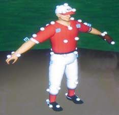 After calibration of each system (Figure 4), the pitcher
threw a series of seven fastballs using a regulation baseball off
a regulation pitcher’s mound. Using positional data from the
camera-based tracking system, real-time three-dimensional
cartoon reconstructions of each pitch were performed (Figure
5). Kinematic parameters were calculated from simultaneous
recordings of position, acceleration, and velocity by the two
systems. The acceleration phase of the pitching cycle was isolated
from each data and maximum acceleration and velocities
compared at the wrist, shoulder, and hand.
After calibration of each system (Figure 4), the pitcher
threw a series of seven fastballs using a regulation baseball off
a regulation pitcher’s mound. Using positional data from the
camera-based tracking system, real-time three-dimensional
cartoon reconstructions of each pitch were performed (Figure
5). Kinematic parameters were calculated from simultaneous
recordings of position, acceleration, and velocity by the two
systems. The acceleration phase of the pitching cycle was isolated
from each data and maximum acceleration and velocities
compared at the wrist, shoulder, and hand.
RESULTS
Results are pictured in Figure 6 and indicate a pitching
acceleration phase lasting 0.022 seconds. The high-speed
motion-tracking camera system was able to capture four data
points during this phase of the pitching cycle. The IMU array
captured 30 data points during this same period. A rapid rise
in elbow extension velocity and humeral internal rotation was
recorded in both systems.
IMU calculations of internal rotation velocity at the shoulder
approximated 12,000 deg/sec. 60.4g’s (591 m/s2) of acceleration
was recorded by the IMU at the shoulder. At the wrist,
80g’s of acceleration (784 m/s2) was recorded at the endpoint of
the acceleration phase of the pitching cycle. Further distally, at
the hand, the IMU array documented greater than 100g’s (980
m/s2) of acceleration.
No statistical comparison between systems was possible
in this early pilot study. Nonetheless, the IMU array recorded
appropriate rises in acceleration and velocity when compared
to the camera-based motion-analysis system. Subjective comparisons
of three-dimensional reconstructions of each pitch
with IMU array data indicated that fine movements of the arm
during the pitching cycle were captured by the IMU array.
DISCUSSION
This proof-of-concept study was designed to validate a
new method of biomechanical analysis using a portable and
wearable set of accelerometers to analyze motion about the
elbow and shoulder during a baseball pitch. A single professional
baseball pitcher underwent simultaneous biomechanical
testing with a traditional “high-speed” motion-analysis
cameras tracking arm position at 180Hz as well as with our
novel IMU array directly measuring arm accelerations and
velocities at 1000Hz.
While one previous study almost 25 years ago attempted
the use of an accelerometer to assess pitching biomechanics,
limits in technology at the time prevented meaningful
results.26 To our knowledge, no further use of accelerometers
to measure pitching has been published, and this study represents
the first productive application of this technology to
this effort.
While statistical comparison was not possible in this small
pilot study, our results indicate that both systems recorded
appropriate rises in acceleration and velocity throughout the
pitching cycle. Subjective comparisons of 3D video reconstructions
indicated that fine movements of the arm during
the pitching cycle were appropriately recorded by the IMU
array.
The IMU array captured almost six times more data during
the acceleration phase of the pitching cycle alone when
compared to the traditional motion-tracking analysis. This
allows a more detailed evaluation of the forces involved in
pitching than what is possible with traditional motion-analysis
cameras. Furthermore, the IMU array directly measured
three-dimensional accelerations and velocities thus avoiding
the possibility of smoothing errors in the process of deriving
acceleration and velocities from position data. Contrary to
previous assumptions regarding acceleration, this initial analysis
demonstrates acceleration within the acceleration phase
to be non-linear (Figure 6). Given this finding, characterization
of forces in the shoulder based on acceleration derived
from traditional technology may be inadequate.
This study presents the first published values of maximal
acceleration in the upper extremity of a professional baseball
player. Moreover, as acceleration is directly proportional to
force, future application of these measurements may allow
greater accuracy in the calculation of forces in the shoulder
and upper arm during the pitching cycle.
Internal rotation velocities at the shoulder during the
acceleration phase of the pitching cycle measured in this subject
approximated 12,000 deg/sec. This is faster (over 15%)
than previously published data10, 14 and could indicate that the
published values of maximum velocity of internal rotation at
the shoulder may be inaccurate and could be refined with a
more complete analysis. Previous calculations of forces in the
upper extremity would also be affected.
IMU arrays are self-contained and allow measurement of
pitching parameters outside of an artificially controlled laboratory
setting. As such, the technology more easily permits
real world and longitudinal studies of the biomechanics of
pitching. Future studies of biomechanics may take place on
an actual playing field or even during a game. We anticipate
that as technology advances and miniaturizes, changes in
biomechanics may be tracked over time, throughout a single
game, or even a career. This would potentially permit quantitative
evaluations and, significant, real-world / real-time
observations of the variation in forces in the pitching arm
as it varies with pitch type, pitcher age, or even with fatigue.
Similarly, changes to training routines and physical conditioning
may be indicated based on a more robust and proactive
monitoring mechanism. Alterations in pitching mechanics
could be evaluated post-operatively and the effects of rehabilitation
quantified.
Moreover, these same analyses could be applied to other
overhead sports such as football or tennis, or even applied to
the lower extremity for biomechanical evaluations outside
of the laboratory in running sports. We hope to attain these
goals with the continued development of this technology.
In conclusion, this pilot study demonstrated the plausible
measurement of the biomechanics of pitching at speeds more
than five times that of traditional tracking cameras. These
wearable IMU’s directly measure velocity and acceleration,
providing the possibility of a new generation of precision in
the measurement of the kinematics and kinetics of pitching.
The measurement mechanism itself more easily allows
measurements outside of controlled laboratory conditions.
The scientific evaluations and real-time, quantifiable observations
that such devices allow have wide-ranging practical and
clinical applications for the overhead throwing athlete including
injury prevention, conditioning / training direction, and
post-operative rehabilitation. We plan to use this system to
study the kinetics and kinematics of different types of pitches
as well including fastballs, curveballs, change-ups, and sliders.
This data would have significant impact on the rehabilitation
and return to pitching of an injured player, as well as provide
objective data on which to introduce new types of pitches to
adolescent players.
Eric Berkson M.D. is a Sports Medicine Fellow at Massachusetts General Hospital.
Ryan Aylward M.S. is a researcher at the MIT Media Laboratory at the Massachusetts
Institute of Technology.
James Zachazewski D.P.T. A.T.C. is the Clinical Director of Sports Medicine at
Massachusetts General Hospital and an Adjunct Assistant Clinical Professor at MGH
Institute of Health Professions.
Joseph Paradiso Ph.D. is an Associate Professor at Massachusetts Institute of
Technology and Sony Career Development Professor of Media Arts and Sciences.
He is also the Co-Director of the Things That Think Consortium at the MIT Media
Laboratory.
Thomas J. Gill, M.D. is an Associate Professor of Orthopaedic Surgery at Harvard
Medical School and Medical Director of the Boston Red Sox.
Address correspondence to:
Thomas J. Gill, M.D.
Massachusetts General Hospital
55 Fruit Street, YAW 3G
Boston, MA 02114
References:
- Tullos HS, King J. Throwing Mechanism in Sports. Orthop Clin North Am 1973;4:709-20.
- Conte S, Requa RK, Garrick JG. Disability days in major league baseball. Am J Sports Med 2001;29(4):431-6.
- Fleisig GS, Kingsley DS, Loftice JW, et al. Kinetic Comparison Among the Fastball, Curveball, Change-up, and Slider in Collegiate Baseball Pitchers. Am J Sports Med 2005.
- Fleisig GS, Andrews JR, Dillman CJ, Escamilla RF. Kinetics of baseball pitching with implications about injury mechanisms. Am J Sports Med 1995;23(2):233-9.
- Rizio L, Uribe JW. Overuse injuries of the upper extremity in baseball. Clin Sports Med 2001;20(3):453-68.
- Chambless KM, Knudtson J, Eck JC, Covington LA. Rate of injury in minor league baseball by level of play. Am J Orthop 2000;29(11):869-72.
- Torg JS, Pollack H, Sweterlitsch P. The effect of competitive pitching on the shoulders and elbows of preadolescent baseball players. Pediatrics 1972;49(2):267-72.
- Andrews J, Wilk K. Shoulder injuries in baseball. In: Andrews J, Wilk K, eds. The Athlete’s Shoulder. New York: Churchhill Livingstone; 1994:369-89.
- Seaver T. The Art of Pitching. New York: Hearst Books; 1984.
- Werner SL, Gill TJ, Murray TA, Cook TD, Hawkins RJ. Relationships between throwing mechanics and shoulder distraction in professional baseball pitchers. Am J Sports Med 2001;29(3):354-8.
- Werner SL, Murray TA, Hawkins RJ, Gill TJ. Relationship between throwing mechanics and elbow valgus in professional baseball pitchers. J Shoulder Elbow Surg 2002;11(2):151-5.
- Escamilla R, Fleisig G, Barrentine S, Zheng N, Andrews J. Kinematic comparisons of throwing different types of baseball pitches. J Appl Biomech 1998;14:1-23.
- Werner SL, Fleisig GS, Dillman CJ, Andrews JR. Biomechanics of the elbow during baseball pitching. J Orthop Sports Phys Ther 1993;17(6):274-8.
- Dillman CJ, Fleisig GS, Andrews JR. Biomechanics of pitching with emphasis upon shoulder kinematics. J Orthop Sports Phys Ther 1993;18(2):402-8.
- Feltner M, Dapena J. Dynamics of the shoulder and elbow joints of the throwing arm during a baseball picth. Int J Sport Biomech 1986;2:235-59.
- Pappas AM, Zawacki RM, Sullivan TJ. Biomechanics of baseball pitching. A preliminary report. Am J Sports Med 1985;13(4):216-22.
- Chen K, Bassett D. The Technology of Accerometry-Based Activity Monitors: Current and Future. Medicine & Science in Sports and Exercise 2005;37(11):S490-S500.
- Williamson R, Andrews BJ. Detecting absolute human knee angle and angular velocity using accelerometers and rate gyroscopes. Med Biol Eng Comput 2001;39(3):294-302.
- Mayagoitia RE, Nene AV, Veltink PH. Accelerometer and rate gyroscope measurement of kinematics: an inexpensive alternative to optical motion analysis systems. J Biomech 2002;35(4):537-42.
- Dejnabadi H, Brigitte J, Aminian K. A New Approach to Accurate Measurement of Uniaxial Joint Angles Based on a Combination of Accelerometers and Gyroscopes. IEEE Transactions on Biomedical Engineering 2005;52(8):1478-84.
- Luinge HJ, Veltink PH. Measuring orientation of human body segments using miniature gyroscopes and accelerometers. Med Biol Eng Comput 2005;43(2):273-82.
- Simcox S, Parker S, Davis GM, Smith RW, Middleton JW. Performance of orientation sensors for use with a functional electrical stimulation mobility system. J Biomech 2005;38(5):1185-90.
- Trost SG, McIver KL, Pate RR. Conducting accelerometer-based activity assessments in field-based research. Med Sci Sports Exerc 2005;37(11 Suppl):S531-43.
- Benbasat A. An Inertial Measurement Unit for User Interfaces [S.M. Thesis]; 2000.
- Benbasat A, Paradiso J. An Inertial Measurement Framework for Gesture Recognition and Applications. In: Wachsmuth I, Sowa T, eds. Gesture and Sign Language in Human-Computer Interaction. London, UK: Springer-Verlag; 2002.
- Hang YS, Lippert FG, 3rd, Spolek GA, Frankel VH, Harrington RM. Biomechanical study of the pitching elbow. Int Orthop 1979;3(3):217-23.
|